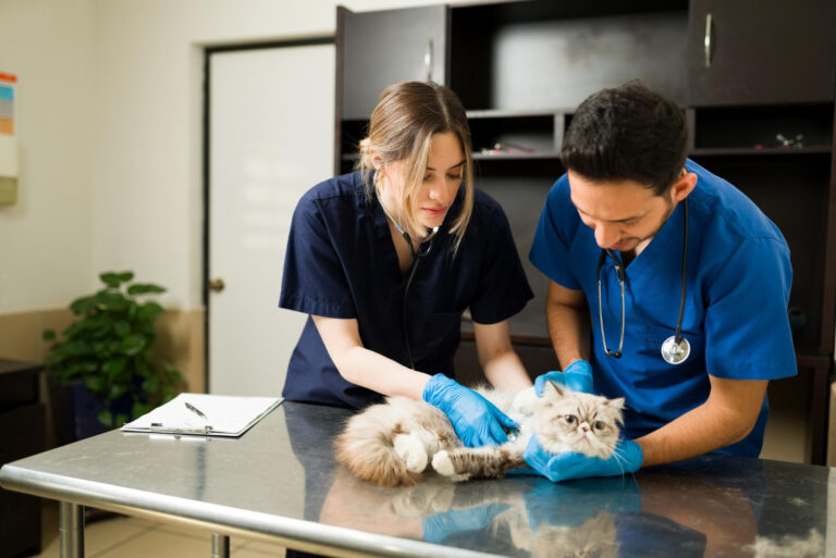

When a pet is feeling unwell, veterinarians often need to look beneath the surface to find the root of the problem. Diagnostic imaging makes this possible without invasive procedures, allowing the care team to examine internal structures and develop an accurate treatment plan. Diagnostic imaging for pets is not a single procedure. It is a category of tests that includes several different technologies, each with its own strengths. X-rays and ultrasound are two of the most frequently used imaging tools in veterinary medicine. Both are safe and effective, but they answer different questions. Knowing how they compare can help pet owners feel more prepared when a veterinarian recommends one of these tests. The right choice depends on the pet’s condition, the area of the body being evaluated, and the information the veterinarian needs to gather.
What Is Diagnostic Imaging for Pets?
Diagnostic imaging refers to a group of non-invasive techniques that allow veterinarians to see inside a pet’s body. These methods are used to examine internal structures and gather the information needed for an accurate diagnosis. The main imaging methods used in veterinary practice include radiography, which is more commonly known as X-ray, as well as ultrasound, CT scans, MRI, real-time X-ray called fluoroscopy, and contrast radiology studies. Each method provides different information, and the choice depends on the pet’s condition and the area of the body being evaluated.
Imaging services are available for a wide range of animals. Veterinary practices and specialty centers offer high-quality imaging services and interpretations for dogs, cats, horses, and some exotic animal species. Whether a pet is recovering from an injury, experiencing digestive problems, or preparing for a procedure, diagnostic imaging helps the veterinary team deliver accurate diagnoses and effective treatment plans. Because these tools are non-invasive, they also reduce stress and discomfort compared with exploratory surgery.
X-Rays: The Most Common Imaging Method in Veterinary Clinics
Radiography, or X-ray imaging, is the most common imaging method used in veterinary clinics. It is often the first step when a veterinarian needs to evaluate a pet quickly. The procedure is painless, non-invasive, and considered very safe for dogs and cats. Digital X-rays in particular use only very low doses of radiation, which makes them a practical option for routine evaluations as well as more complex cases. In addition to small animal practices, portable X-ray machines are used for large animals such as horses, allowing imaging to be performed wherever the animal is located.
X-rays provide valuable information about the body’s internal structures and are often used to investigate signs of illness or injury. Because the images are captured quickly and the technology is widely available, X-rays serve as an efficient starting point in many diagnostic workups. If the initial images do not provide enough detail, the veterinarian may recommend additional imaging methods to gain a clearer understanding of the pet’s condition.
Ultrasound: A Safe Way to See Inside Your Pet
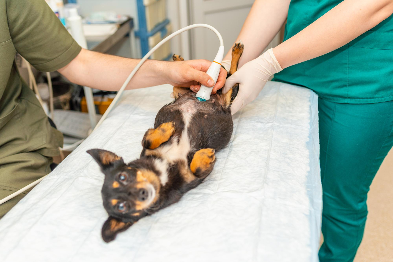
Ultrasound uses state-of-the-art imaging technology to allow veterinarians to safely see inside a pet without invasive procedures. For many pet owners, this is one of the most reassuring aspects of the test: there is no surgery, no incision, and no lengthy recovery period. Ultrasound is particularly valuable for abdominal and thoracic imaging, including echocardiography, which evaluates the heart. The technology gives the veterinary team a detailed look at internal structures while keeping the pet comfortable throughout the examination.
Ultrasound services are offered in a variety of settings. Some specialty practices were created specifically to improve access to veterinary specialists, recognizing that not every community has a specialist nearby. Mobile ultrasound services and teleradiology also extend the reach of expert interpretation, allowing general practice veterinarians to consult with specialists without sending the pet to another facility.
Many ultrasound providers are Fear Free Certified, meaning the team has been trained to reduce stress for both the pet and the owner. Owners are often encouraged to stay with their pet during the appointment, which typically lasts about one hour. There are no extended wait times to get into a specialty hospital, and there is no need to drop off a frightened pet. This approach helps anxious animals remain calm and gives owners the chance to ask questions throughout the process.
How Veterinarians Choose Between Ultrasound and X-Ray
There is no single imaging test that fits every pet. Veterinarians choose the method based on the pet’s history, the results of the physical examination, and the specific information they need to gather. X-rays are often the starting point because they are the most common and most accessible imaging method in veterinary clinics. If the X-rays reveal an area that needs closer evaluation, or if the clinical signs point toward a soft tissue or heart concern, an ultrasound may be the next step.
In some cases, the veterinarian may recommend more than one imaging method. The full range of veterinary imaging tools includes radiography, CT scans, MRI, real-time X-ray fluoroscopy, ultrasound, and contrast radiology studies. These methods are sometimes used together to deliver the most accurate diagnosis and the most effective treatment plan. A pet with a complex condition may need an X-ray for a broad view and an ultrasound for a detailed look at a specific organ. In other situations, a referral to a specialty imaging center may be the best path forward, particularly when advanced technology or specialist interpretation is required.
Both X-rays and ultrasound are considered safe, and both avoid the need for surgery. Imaging is usually recommended only when the veterinarian believes the results will meaningfully change how a pet is treated. In many cases, the information gained from these tests helps the team act more quickly and with greater confidence.
The Role of Specialist Interpretation
High-quality images are only part of the equation. Accurate interpretation is just as important, which is why many veterinary practices rely on specialists. Teleradiology services allow digital images to be transmitted to a specialist who can review them and provide a detailed report. This is especially valuable in areas where access to veterinary specialists is limited. Imaging centers that offer mobile ultrasound and teleradiology help bridge that gap, so pets in smaller communities can still benefit from specialist-level care without traveling long distances.

What to Expect During an Imaging Appointment
Imaging appointments are designed to be as comfortable as possible for both the pet and the owner. In many ultrasound practices, there is no need to drop off a frightened pet. Instead, owners are encouraged to stay with their pet during the appointment, which typically lasts about one hour. Fear Free Certified teams work to reduce stress throughout the visit, and the focus remains on keeping the animal calm and relaxed. This approach is especially helpful for pets that become anxious in unfamiliar settings.
X-ray appointments tend to be shorter and may not require the same level of preparation. The procedure itself is painless and non-invasive, and digital X-rays use only very low doses of radiation. For large animals like horses, portable X-ray machines allow imaging to be performed in a barn or field setting. After the images are captured, they are reviewed and interpreted by the veterinary team. If teleradiology is available, the images may also be sent to a specialist for review, ensuring that the pet benefits from expert interpretation even when the specialist is located elsewhere.
Frequently Asked Questions
Are X-rays safe for my pet?
Yes. X-rays are painless, non-invasive, and considered very safe for dogs and cats. Digital X-rays in particular use only very low doses of radiation. Because the images are captured quickly, most pets do not need sedation, and the procedure can often be completed during a regular office visit. Your veterinarian will only recommend an X-ray when the information it provides is expected to help with the diagnosis and treatment plan.
Does an ultrasound hurt my pet?
No. Ultrasound technology allows veterinarians to safely see inside your pet without invasive procedures. Many providers are Fear Free Certified to reduce stress for the pet, and owners are often encouraged to stay with their pet during the appointment. A typical ultrasound visit lasts about one hour, giving the veterinary team time to examine the area of concern while keeping the animal comfortable.
Why might my veterinarian recommend both X-rays and ultrasound?
Different imaging methods provide different information. X-rays are the most common imaging method in veterinary clinics and are often the first test performed. Ultrasound offers a safe, non-invasive way to examine internal structures, which makes it a useful follow-up when more detail is needed. In complex cases, veterinarians may combine radiography, ultrasound, CT, MRI, fluoroscopy, and contrast studies to reach the most accurate diagnosis.
What other diagnostic imaging options are available for pets?
Beyond X-rays and ultrasound, veterinary diagnostic imaging includes CT scans, MRI, real-time X-ray fluoroscopy, and contrast radiology studies. Mobile ultrasound services and teleradiology also help expand access to veterinary specialists for pets in communities without a dedicated specialty hospital. Your veterinarian can explain which imaging method is appropriate for your pet’s specific condition and what the results will reveal about the next steps in care.