Veterinary diagnostic imaging is one of the most frequently used categories of diagnostic testing in both small and large animal hospitals throughout the world. When a veterinarian recommends an X-ray, ultrasound, CT scan, or MRI, it is natural to have questions about why the test is needed and what it will involve. In 2026, the imaging tools available in veterinary medicine continue to improve, giving veterinarians a clear view of what is happening inside a pet’s body. This article covers the basics of veterinary diagnostic imaging, the common modalities, and what pet owners should expect during the process.
The goal of diagnostic imaging is to help pets receive accurate diagnoses and effective treatment. Imaging studies are used across nearly every area of veterinary medicine, from routine abdominal checks to complex neurologic cases. Understanding how these tools work can help pet owners feel more confident when their veterinarian suggests an imaging study.
What Is Veterinary Diagnostic Imaging?
Veterinary diagnostic imaging is a specialized branch of veterinary medicine that uses various imaging techniques to help diagnose and monitor animal diseases. These techniques include X-rays, ultrasound, CT scans, MRI, and other imaging modalities that provide detailed views of an animal’s internal structures. These images can help veterinarians identify fractures, tumors, organ abnormalities, and other medical conditions, supporting accurate diagnosis and effective treatment planning.
Imaging is not limited to one body system. Veterinary radiology education covers radiographs, ultrasound, CT, and MRI, with common regions including the thorax, abdomen, musculoskeletal system, and neurologic system. Both canine and feline patients are regularly imaged, which means dogs and cats of all ages can benefit from these diagnostic tools. By creating a clear picture of internal anatomy, imaging reduces guesswork and helps the care team explain findings to pet owners in concrete terms.
Common Imaging Modalities in Veterinary Medicine
The most common imaging modalities in veterinary medicine are radiography, ultrasonography, computed tomography, and magnetic resonance imaging. Each one serves a different purpose, and veterinarians choose the technique based on the body region and the suspected condition.
| Modality | What It Produces | Common Uses |
|---|---|---|
| Radiography (X-rays) | Two-dimensional images of internal structures | Abdominal and musculoskeletal radiographs |
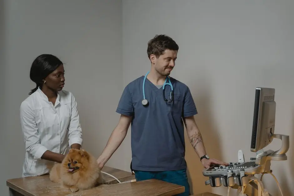
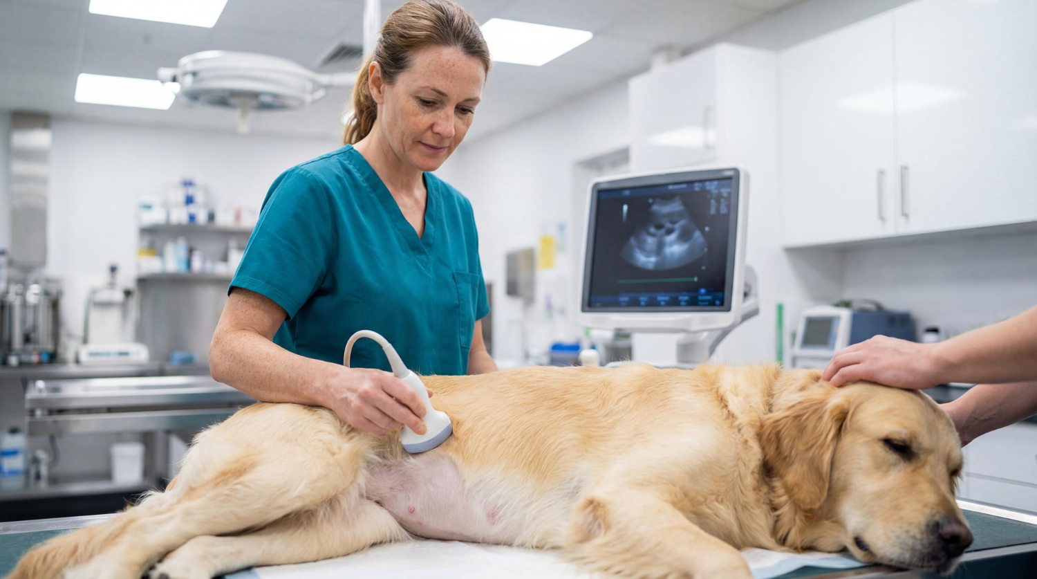
| Ultrasonography | Real-time images of soft tissues using sound waves | Abdominal and thoracic evaluation, echocardiograms |
| Computed Tomography (CT) | Detailed cross-sectional images | Thorax, abdomen, musculoskeletal, and neurologic imaging |
| Magnetic Resonance Imaging (MRI) | Highly detailed images of soft tissues | Neurologic and soft tissue evaluation |
Radiographs, commonly known as X-rays, are among the most frequently used imaging tools in veterinary practice. Many veterinary hospitals use abdominal and musculoskeletal radiographs as part of routine and emergency care. Ultrasonography uses sound waves to examine soft tissues and organs, while specialized ultrasound exams such as echocardiograms focus on the heart. CT and MRI are advanced imaging techniques that provide greater detail and can be especially valuable when evaluating the thorax, abdomen, musculoskeletal system, or neurologic system.
How Imaging Is Used Across the Body
Veterinary diagnostic imaging is used across the full range of patient care. Radiographs are routinely ordered for the abdomen and musculoskeletal system, helping veterinarians evaluate the stomach, intestines, bones, and joints. Ultrasound is commonly used for the thorax and abdomen, where it can reveal changes in organs such as the liver, kidneys, and spleen, as well as the heart during an echocardiogram. CT and MRI are frequently reserved for the thorax, abdomen, musculoskeletal system, and neurologic system, including cases that involve the brain and spinal cord.
The teaching model in veterinary radiology reflects these same categories, with training organized around the thorax, abdomen, musculoskeletal system, and neurologic system. That structure ensures veterinarians are prepared to image any part of the body and to recognize both normal anatomy and disease.
Why Diagnostic Imaging Matters for Pet Health
Diagnostic imaging is one of the most frequently used categories of diagnostic testing in both small and large animal hospitals throughout the world. That frequent use is driven by a simple reality: many medical conditions cannot be confirmed through a physical examination alone. Imaging provides the detailed pictures that allow veterinarians to see inside the body without the need for exploratory surgery.
The benefits are significant. Fractures, tumors, organ abnormalities, and other medical conditions can be identified clearly, which enables accurate diagnosis and effective treatment plans. For a pet that is limping, coughing, vomiting, or showing neurologic signs, imaging often gives the veterinarian the information needed to recommend the right next step. Imaging is also valuable for monitoring chronic conditions, allowing veterinarians to compare images over time and adjust treatment as the patient’s condition changes.
Advanced Imaging Technology in 2026
Veterinary hospitals today have access to advanced imaging technologies that help veterinarians evaluate a wide range of conditions with greater accuracy. Modern diagnostic imaging may include digital radiographs, ultrasound, CT scans, and MRI, depending on the pet’s symptoms and the type of information needed.
For pet owners, the practical takeaway is that veterinary imaging in 2026 is more capable than ever. High-quality radiographs and ultrasound are commonly used for many diagnostic needs, while CT and MRI can provide more detailed views of complex conditions. These technologies can help veterinary teams identify abnormalities, confirm diagnoses, and develop more informed treatment plans for pets.
The Role of Board-Certified Veterinary Radiologists
Behind many veterinary imaging studies is a specialist known as a board-certified veterinary radiologist. These veterinarians complete advanced training in diagnostic imaging and are certified by the American College of Veterinary Radiology. Some veterinary practices work directly with board-certified veterinary radiologists, while others use teleradiology services, where medical images are securely sent to a radiologist for professional interpretation.
Teleradiology allows veterinary teams to access specialized imaging expertise without requiring pets to travel long distances. Depending on the practice and the type of imaging needed, radiologists may interpret digital radiographs, ultrasound studies, CT scans, and other diagnostic images. Their expertise can help veterinarians better understand complex findings, confirm diagnoses, and make informed treatment recommendations.
Keeping Pets Calm During Imaging Appointments
Frequently Asked Questions
The questions below are among those pet owners most often ask about veterinary diagnostic imaging.
What is the difference between radiography, ultrasound, CT, and MRI?
Radiography, or X-ray, is commonly used for abdominal and musculoskeletal imaging. Ultrasound uses sound waves to evaluate soft tissues and organs in the abdomen and chest. CT creates detailed cross-sectional images useful for the thorax, abdomen, musculoskeletal system, and neurologic system. MRI provides highly detailed soft tissue images and is often used for neurologic cases. Your veterinarian selects the best option based on the region being examined and the suspected condition.
What conditions can veterinary diagnostic imaging detect?
Veterinary diagnostic imaging helps identify fractures, tumors, organ abnormalities, and other medical conditions. Because it produces detailed pictures of internal structures, it is useful for evaluating the thorax, abdomen, musculoskeletal system, and neurologic system. Common imaging studies include abdominal and musculoskeletal radiographs as well as ultrasound, CT, and MRI. The images allow veterinarians to confirm a diagnosis and build an effective treatment plan for dogs and cats.
Can I stay with my pet during an imaging appointment?
In many cases, yes. Some veterinary diagnostic centers encourage owners to stay with their pet for the duration of a one-hour appointment rather than dropping the pet off. These practices often use Fear Free certified techniques to reduce stress for pets and owners. However, protocols vary by hospital and by the type of imaging being performed. Ask your veterinary team ahead of time about their policy for owner presence during imaging.
What is a board-certified veterinary radiologist?
A board-certified veterinary radiologist is a specialist who has completed advanced training in veterinary diagnostic imaging. These specialists are trained to interpret imaging studies such as X-rays, ultrasound, CT scans, and MRI. Some veterinary practices may use teleradiology, which allows diagnostic images to be securely sent to a veterinary radiologist for expert review. This gives primary care veterinarians access to specialized imaging expertise without requiring the pet to travel to another facility. Their interpretation can help support accurate diagnosis and informed treatment planning.
Conclusion
Veterinary diagnostic imaging gives veterinarians valuable insight into a pet’s internal health, helping identify conditions that may not be visible during a physical examination. From X-rays and ultrasound to CT and MRI, each imaging method serves a specific purpose in diagnosis and treatment planning. If your pet has been experiencing unusual symptoms or your veterinarian recommends diagnostic imaging, contact Wareham Animal Hospital to discuss your pet’s needs and learn how the right imaging approach can support timely, informed care.