
When your dog or cat suddenly starts coughing, breathing with their mouth open, or acting unusually tired, it can be alarming. These signs are often more than everyday discomfort. They may point to underlying conditions affecting the lungs or heart that require immediate veterinary attention.
According to the American Veterinary Medical Association (AVMA), respiratory and cardiovascular diseases are among the leading causes of illness in household pets. Early recognition of symptoms such as coughing, labored breathing, and lethargy can make a critical difference in treatment outcomes. This is why veterinary teams rely on tools like thoracic ultrasound, a non-invasive way to see what is happening inside your pet’s chest.
These symptoms can be confusing and sometimes subtle. A dog’s cough may sound like a simple irritation, but it could be linked to heart disease. A cat’s open-mouth breathing may look like panting, but it can signal a serious emergency. Lethargy, often dismissed as “just being tired,” may be an early red flag for infections or metabolic disorders. Understanding these signs and acting quickly can help save lives.
Key Takeaways
- Coughing, open-mouth breathing, and lethargy often signal underlying respiratory or heart disease.
- Thoracic ultrasound is a safe and effective tool to evaluate the lungs and heart in pets.
- Early veterinary intervention improves the chances of recovery and reduces complications.
What Is a Thoracic Ultrasound?

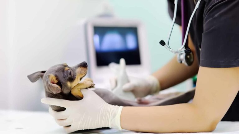
A thoracic ultrasound is an imaging test that uses sound waves to create detailed pictures of the heart, lungs, and surrounding tissues inside your pet’s chest.
Unlike X-rays, it does not involve radiation, making it a gentle and safe choice. This procedure is especially valuable when pets show signs of respiratory distress. It allows veterinarians to evaluate conditions like pleural effusions (fluid around the lungs), heart enlargement, or tumors. Because it provides real-time images, it also helps guide treatment decisions, such as whether fluid should be drained or additional testing is needed.
The Core Symptoms That Signal Trouble
Coughing
Coughing is one of the most common signs that something is wrong with a pet’s respiratory or cardiovascular system. While the occasional cough may simply be the body’s way of clearing irritants, persistent or worsening coughing deserves attention.
Causes of Coughing in Pets:
- Heart Disease: Both dogs and cats may cough due to heart failure. The cough is often dry but can sometimes produce frothy fluid.
- Respiratory Infections: Viral or bacterial infections can lead to bronchitis or pneumonia.
- Asthma (in Cats): Feline asthma often presents with wheezing and repetitive coughing fits.
- Allergies and Irritants: Dust, smoke, or chronic bronchitis may trigger frequent coughing.
- Parasitic Infections: Heartworm and lungworm are known culprits.
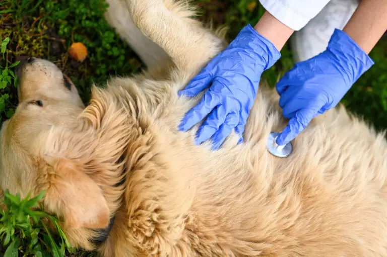
Veterinarians evaluate coughing by combining a physical exam with diagnostic tools like X-rays, bloodwork, and ultrasounds. Dogs may show lethargy and soft coughs, while cats can exhibit wheezing that resembles human asthma.
Open-Mouth Breathing
Open-mouth breathing is far more concerning than panting. Panting helps dogs cool down after exercise, but open-mouth breathing, especially in cats, almost always signals distress.
Possible Causes:
- Asthma or Airway Inflammation narrows the passages for airflow.
- Congestive Heart Failure leading to fluid buildup in the lungs.
- Obstructions such as tumors or foreign objects blocking breathing pathways.
- Severe Infections like pneumonia.
Emergency Signs That Require Immediate Veterinary Care:
- Pale or bluish gums (oxygen deprivation).
- Collapse or fainting during episodes.
- Wheezing or gasping that worsens with time.
When veterinarians encounter these symptoms, thoracic ultrasound can quickly help confirm whether the problem lies in the heart, lungs, or surrounding chest cavity.
Lethargy
Lethargy is more than tiredness. It is a state where pets lose their usual interest in food, play, and daily activity. While it might seem like a minor change, lethargy can be a key symptom of systemic illness.
Common Causes of Lethargy:
- Infections such as bacterial, viral, or fungal diseases.
- Respiratory Illnesses that reduce oxygen flow, leaving pets exhausted.
- Gastrointestinal Disorders such as inflammatory bowel disease.
- Metabolic Diseases, including diabetes, hypothyroidism, or kidney failure.
A lethargic pet needs a thorough veterinary examination. Blood tests, X-rays, and ultrasounds are often recommended to pinpoint the cause. Because lethargy overlaps with many conditions, identifying the root problem quickly is critical.
Why Thoracic Ultrasound Matters
When a pet shows signs like coughing, open-mouth breathing, or lethargy, it can be difficult to pinpoint the exact cause.
For example, a cough might come from heart disease, an infection, or even an obstruction. On the surface, these conditions look very similar, but each requires a very different type of care. Thoracic ultrasound provides the clarity veterinarians need to understand what is truly happening inside a pet’s chest.
With this non-invasive tool, veterinarians can see in real time if fluid has built up. If the heart is enlarged, or if there is a mass pressing on the lungs. That information is critical because it helps guide treatment without unnecessary delays.
Advantages of Thoracic Ultrasound
- Non-invasive and painless for pets.
- Produces real-time, detailed images of the heart and lungs.
- Helps distinguish between conditions that appear alike.
- Can guide treatments such as draining fluid from the chest.
- Complements radiographs to create a complete picture of the chest.
Used alongside other diagnostic tools, thoracic ultrasound allows veterinarians to make accurate and timely decisions that can improve a pet’s prognosis and overall quality of life.
“Thoracic ultrasound gives us the answers that X-rays alone can’t. It helps us see what’s really happening in a pet’s chest so we can act quickly and give them the care they need.”
— Dr. Alberto L. Fernandez, Wareham Animal Hospital
The Thoracic Ultrasound Procedure
Preparation: Before the procedure, your pet’s medical history and prior diagnostic results are reviewed. Sedation may be used if a pet is anxious, but many relax naturally during the process.
How It Works: A veterinarian applies gel to the chest and uses a handheld probe to capture images. The sound waves pass into the body and bounce back, creating live images of organs and tissues.
Interpreting Results: The images are analyzed alongside other tests and the pet’s clinical signs. This helps identify conditions such as heart enlargement, pleural effusions, pneumonia, or tumors. Results are then used to guide an appropriate treatment plan.
Current Market Trends in Veterinary Diagnostics
In recent years, there has been a growing demand for advanced, non-invasive diagnostic imaging in veterinary medicine. Thoracic ultrasound has become more widely available as pet owners increasingly seek accurate, fast, and low-stress diagnostic methods.
According to industry reports, diagnostic imaging services in veterinary practices are expanding, with ultrasound use rising by over 20% in the past five years. This trend reflects both improved technology and the increasing awareness of pet owners about the importance of early detection. For conditions involving the chest, thoracic ultrasound is now considered an essential tool rather than an optional procedure.
Why Choose Wareham Animal Hospital
Wareham Animal Hospital understands how stressful it can be when your pet shows signs like coughing, open-mouth breathing, or lethargy.
Our veterinary team is dedicated to providing compassionate and thorough care using the most advanced diagnostic tools available. We offer thoracic ultrasound as part of our commitment to accurate, timely diagnosis. Our team takes the time to explain each step of the process, answer your questions, and ensure that both you and your pet feel supported throughout their care.
Choosing Wareham Animal Hospital means choosing a hospital where your pet’s health and comfort always come first.
The Bottom Line
Coughing, open-mouth breathing, and lethargy are not symptoms to take lightly. These signs can point to serious respiratory or cardiovascular issues that require urgent attention. Thoracic ultrasound provides a safe and effective way to look inside your pet’s chest and uncover the real cause of their illness. Early diagnosis not only improves treatment success but also helps ensure your beloved companion has the best chance at recovery.
Read more of our related blogs here.
Frequently Asked Questions
1. Is thoracic ultrasound safe for pets?
Yes. Thoracic ultrasound is a non-invasive, painless, and radiation-free procedure that is safe for both dogs and cats.
2. Do pets need sedation for a thoracic ultrasound?
Not always. Most pets tolerate the procedure well. Sedation may only be used if a pet is anxious or unable to stay still.
3. How long does the procedure take?
The average thoracic ultrasound takes about 20–40 minutes, depending on the complexity of the case.
4. What conditions can thoracic ultrasound detect?
It can reveal heart enlargement, fluid around the lungs, tumors, infections, and structural abnormalities in the chest.
5. How soon will I get results?
In most cases, veterinarians can discuss preliminary findings immediately after the procedure and provide a full interpretation shortly thereafter.