
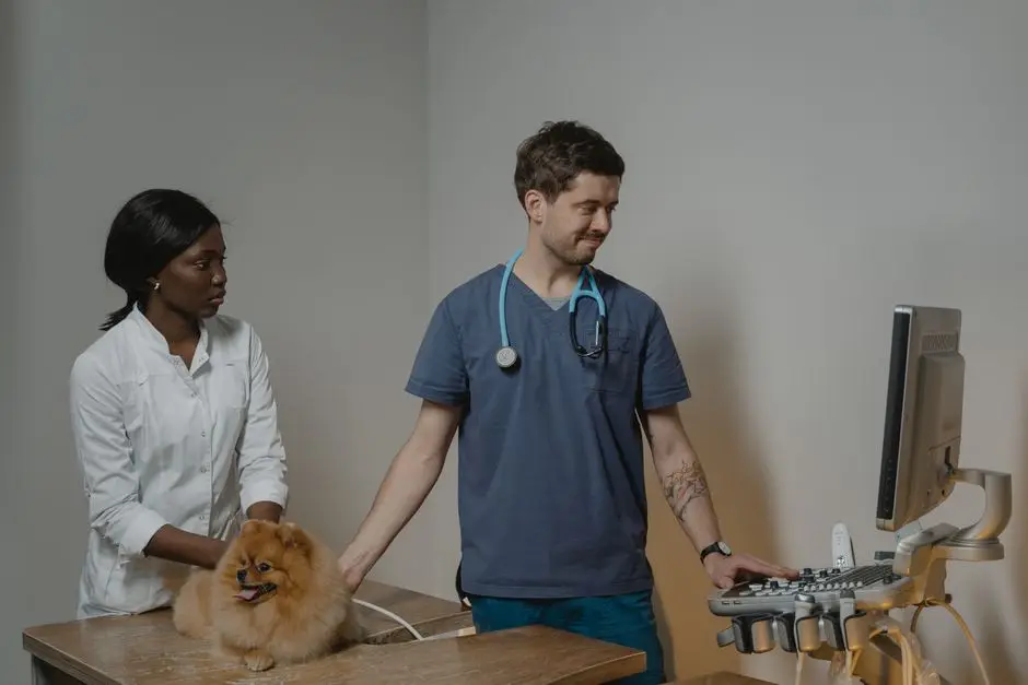
A recommendation for a heart ultrasound can catch a pet owner off guard. The request often arrives after a routine exam, a follow-up visit, or a conversation about a possible heart condition, and it comes with a name that sounds more intimidating than the procedure itself. A heart ultrasound for pets, also called an echocardiogram, is a diagnostic study that gives your veterinarian a detailed look at how your pet’s heart is built and how well it is working. Understanding what the test does, what it cannot do, and how it differs from other heart tests makes the next steps easier to approach with confidence.
What a Heart Ultrasound Actually Is
An echocardiogram is a type of ultrasound for dogs and cats used to evaluate heart function and detect abnormalities. It uses the same basic technology as the ultrasound machines used for other parts of the body, but the probe is aimed at the heart rather than the abdomen or other organs.
Cardiac ultrasounds are described as detailed studies that can closely assess the heart and its surrounding structures. That includes the pericardial sac, the sac that surrounds the heart. A heart ultrasound can help evaluate the condition and function of a pet’s heart and search for abnormalities, which is why it is often used when a veterinarian wants more than a general impression of cardiac health.
Ultrasound in general is described as a highly useful tool when evaluating heart conditions, internal organs, cysts, and tumors. The cardiac version narrows that focus to the heart itself. Targeted veterinary ultrasounds and echocardiograms help answer specific diagnostic questions rather than producing a broad survey of the whole body.
Why Your Veterinarian May Recommend One
For dogs or cats with a heart condition, a veterinarian may recommend an echocardiogram to help find out whether the pet will require heart-related care. That is the core reason behind most referrals for the study. Your veterinarian is not ordering the test simply to gather data. The goal is to answer a question that affects what happens next for your pet.
Evaluating Heart Function
A heart ultrasound can evaluate the condition and function of the heart. Function matters because a heart can look normal in size while still not moving blood the way it should. The study gives the veterinarian a view of how the heart is working as a pump, not just how large it appears to be.
Examining Structure and Surrounding Tissue
Cardiac ultrasounds examine the heart and its surrounding structures, including the pericardial sac. This means the veterinarian can look at how the heart is put together and check the tissue immediately around it. Structural detail of this kind is one of the main reasons the study is chosen over other imaging.
Answering a Specific Diagnostic Question
Targeted veterinary ultrasounds and echocardiograms help answer specific diagnostic questions. Your veterinarian may already have a working theory based on an exam, a blood test, or another imaging study. The echocardiogram is how that theory gets tested with a direct look at the heart.

Heart Ultrasound, X-Ray, and ECG: What Each One Shows
These tests are sometimes confused with one another because they all involve the heart. They are not interchangeable, and each one answers a different question. The table below summarizes the distinction as described in veterinary sources.
| Test | What it shows |
|---|---|
| X-ray | The size, shape, and position of the heart |
| ECG | The heart’s electrical rhythm |
| Heart ultrasound (echocardiogram) | The internal structure and function of the heart, including how the heart is built and surrounding structures such as the pericardial sac |
The clearest way to hold these apart is this: a heart ultrasound shows how the heart is built, while an ECG shows its electrical rhythm. Those are different kinds of information. A rhythm can be irregular while the structure looks unremarkable, and a structural problem can exist without an obvious rhythm disturbance.
The relationship between X-ray and ultrasound is similar. X-rays show the size, shape, and position of the heart, while ultrasound allows the veterinarian to see the internal structure and function of the heart. That is a difference in what each method reveals, not a ranking of one over the other. Your veterinarian selects the study, or the combination of studies, that fits the question being asked.
What the Results Help Your Veterinarian Decide
Once the study is complete, the findings feed into a plan. For pets with a heart condition, the echocardiogram helps the veterinarian determine whether heart-related care will be needed. From there, the conversation usually turns to monitoring, follow-up visits, or other diagnostics that build on what the ultrasound showed.
It is important to keep expectations realistic. An echocardiogram is a diagnostic tool, not a treatment. It produces information. What your veterinarian does with that information depends on the individual pet, the findings, and the overall clinical picture. No single test answers every question about a pet’s heart, which is why the echocardiogram is usually one part of a broader evaluation rather than the entire plan.
One detail worth confirming directly with your veterinary team is cost. Prices for veterinary imaging vary by practice, region, and the specifics of the study, and this article does not quote figures. Ask the hospital for an estimate before the appointment so there are no surprises.

Questions to Ask Before the Appointment
You do not need to become a cardiology expert to be an informed advocate for your pet. A short list of questions can make the appointment more productive and help you understand what is happening.
- What specific question is this ultrasound meant to answer?
- Who will perform the study, and where will it be done?
- Does my pet need any preparation beforehand?
- How will the results change my pet’s care plan?
- Will we need follow-up imaging, and if so, when?
- What is the estimated cost, and what does it include?
- Should I expect a referral to another provider?
Some of these answers depend on the individual practice and the individual pet. Preparation requirements, sedation decisions, procedure length, and referral practices are not the same everywhere, and they are not something you should assume. The most reliable source is the veterinary team handling your pet’s case. Write your questions down before the visit, because it is easy to forget them in the moment.

Finding This Service Locally
Echocardiography is not offered at every veterinary clinic, so it helps to know where the service is available near you. Wareham Animal Hospital is a full-service veterinary practice in Wareham, Massachusetts, serving pet owners in Wareham and nearby southeastern Massachusetts communities including Fall River, Mashpee, Taunton, Lakeville, Berkley, Plymouth, Duxbury, Bridgewater, Falmouth, and New Bedford. The hospital’s services include echocardiography along with abdominal and thoracic ultrasound, surgery, emergency care, and personalized wellness plans. If your pet’s veterinarian has recommended a heart ultrasound, the care team can walk you through what the study involves for your individual dog or cat.
Frequently Asked Questions
Is a heart ultrasound the same as an ECG?
No. They are different studies. A heart ultrasound shows how the heart is built, while an ECG shows the heart’s electrical rhythm. Your veterinarian may use both because each one answers a separate question about cardiac health. If you are unsure which test has been recommended for your pet, ask the veterinary team to clarify before the appointment.
Can an X-ray show the same information as a heart ultrasound?
They reveal different things. X-rays show the size, shape, and position of the heart, while ultrasound allows the veterinarian to see the internal structure and function of the heart. A radiograph gives a silhouette view and an echocardiogram gives a look inside. Your veterinarian chooses based on the diagnostic question being asked.
Is a heart ultrasound the same as an abdominal ultrasound?
The technology is similar, but the focus differs. A cardiac ultrasound examines the heart and its surrounding structures, including the pericardial sac. General ultrasound is described as useful for evaluating heart conditions along with internal organs, cysts, and tumors. An echocardiogram is a targeted study aimed specifically at the heart rather than a broad survey.
What should I ask my veterinarian before scheduling?
Ask what question the study is meant to answer, who performs it, whether any preparation is needed, and how the findings will shape your pet’s care. Cost is another fair question, since imaging prices vary by practice and region. Your veterinary team can give you details specific to your pet and the hospital.